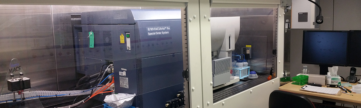
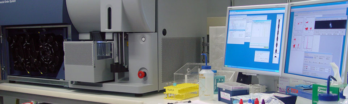
How Cytometers Work (Basic Operation of a Cell Sorter)
 Click on the image for a larger view.
Flow cytometers utilize a sophisticated array of lasers,
optics, fluidics and electronic detectors to measure light
scatter and/or fluorescence emission from cells in purified
suspension that are hydrodynamically focused to a single cell
wide laminar flow column. The fluid column passes through
laser light focused to a beam a few cell diameters across.
Laminar flow permits laser interrogation of the cells in
single file, one cell at a time. As each cell passes through
the laser light it emits scattered light in all directions.
Light scatter measured at 90 degrees from incidence (right or
orthogonal scatter) corresponds to the quantity and light
diffractive quality of granular structures inside a cell, and
light scatter measured at 180 degrees (forward scatter) approximately corresponds to cell size. These parameters
alone are sufficient to distinguish numerous cell types and to
exclude dead cells, aggregates and cell debris from cells of
interest.
If the cells have been stained with fluorochromes
(or transfected to express fluorogenic activity - GFP) they
will also emit fluorescence intensities at levels which
directly correspond to the density of fluorochrome on or
within the cell. Notably, any cell structure for which an
antibody or other ligand probe is available may be
characterized with respect to surface or intracellular
density by fluorescence emission. Any gene whose expression
can be coupled to a fluor such as GFP can identify the cell
in which it is expressed and be further characterized
or sorted on that basis. Further thanks to steering optics and
wavelength filters which retrieve light of various
wavelengths and guide them to separate detectors, fluorescence intensities may be measured at several
different wavelengths simultaneously for each cell, allowing
multiple antigens to be measured.
In cytometers capable of
sorting, the hydrodynamically focused cell stream is broken
into uniform-sized droplets by axial vibration of the
nozzle. A drop-charging signal is triggered by an electronic
system that processes the fluorescent and scatter signals
received from cells and determines the appropriate charge to
give each cell. Only two populations can be retrieved since
the uncharged cells go to waste. Some cytometers have
overcome this limitation and can now separate up to four
populations by producing four side streams by varying the
charge applied to the drop. See U.S. Patent 5,483,469,
January 9, 1996, to inventor Ger Van den Engh. |